Die Röntgenvideographie in der Bewegungsforschung
Bereits 1981 hat Martin S. Fischer bei Jean-Pierre Gasc in Paris die Röntgenvideographie kennengelernt und ab 1988 am Institut für den Wissenschaftlichen Film in Göttingen (IWF Wissen und Medien GmbH) Bewegungen von Säugetieren mit Hilfe dieser Technik systematisch untersucht. Dort stand ein uniplanares Phillips-System zur Verfügung mit einem Bildverstärker von 200 mm x 153 mm Durchmesser und einer Arritechno 35 mm-Filmkamera mit einer Aufnahmegeschwindigkeit von 150 Bildern pro Sekunde. Die Daten wurden zunächst direkt von den Filmrollen aus analysiert. In einem ersten gemeinsamen DFG-Projekt mit Hanns Ruder vom Institut für theoretische Physik der Universität Tübingen wurde die Kinematik und Dynamik der Fortbewegung sowie das Klettern von Klippschliefern erforscht.


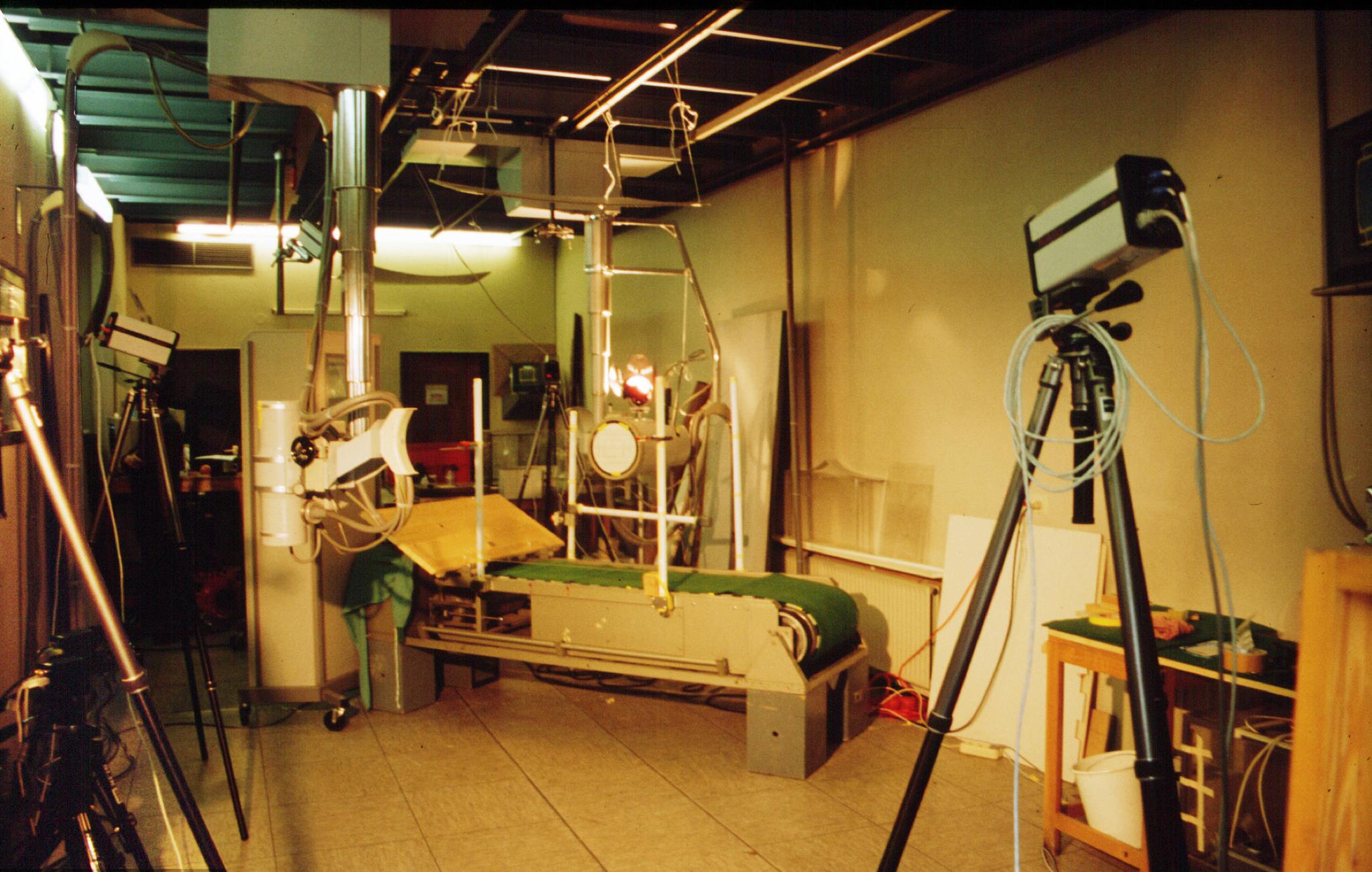
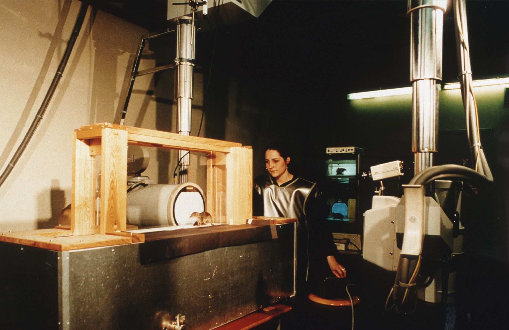
Röntgenvideographie am IWF in Göttingen: selbstkonstruierte Laufbänder, hochmotivierte Forscherinnen und Forscher mit viel Geduld und Improvisationstalent sowie wochenlange Habituation der Tiere sind bis heute unerlässlich für das Gelingen der Studien, rechts: Nadja Schilling bei Aufnahmen für ihre Diplomarbeit zur Fortbewegung des Spitzhörnchens (Tupaia glis) 1997.
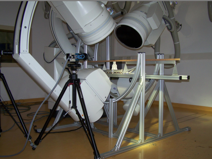
Mit der Berufung von Martin S. Fischer auf den Lehrstuhl für Spezielle Zoologie und Evolutionsbiologie der Universität Jena und der Einrichtung des DFG-Innovationskollegs „Bewegungssysteme“, gemeinsam mit Reinhard Blickhan (Institut für Sportwissenschaften), Hans-Christoph Scholle (Klinik für Unfall-, Hand- und Wiederherstellungschirurgie) und Klaus Zimmermann (FG Technischer Maschinenbau, TU Ilmenau) wurde in Thüringen ein Zentrum für Bewegungsforschung etabliert, das bis heute die evolutionsbiologische und biomechanische Grundlagenforschung mit der Anwendung in den Ingenieurwissenschaften und in der medizinischen Präventionsforschung in weltweit einzigartiger Weise verbindet. Dieses Netzwerk, inzwischen längst über die Grenzen Thüringens hinaus gewachsen, trägt bis heute und bildet seit mehr als 20 Jahren den Nucleus für Forschungsaktivitäten in verschiedenste Richtungen. Hunderte von Publikationen in Zeitschriften und Büchern sind seitdem entstanden und viele Wissenschaftlerinnen und Wissenschaftler haben hier ihre Laufbahn begonnen. Gefördert wurde die Bewegungsforschung in Jena-Ilmenau durch die Deutsche Forschungsgemeinschaft (DFG), durch das Bundesministerium für Bildung und Forschung (BMBF), durch Mittel des Landes Thüringen sowie durch ausländische Institutionen, z.B. in Form von Stipendien für Gastwissenschaftlerinnen und Gastwissenschaftler, die regelmäßig Teil der hiesigen Forschergruppe werden. Im Rahmen einer solchen Großförderung durch das BMBF (Projekt „InspiRat - Entwicklung eines bionisch inspirierten Kletterroboters für die externe Inspektion linearer Strukturen“, Partner: Martin S. Fischer, Hartmut Witte, Stanislav N. Gorb, Andreas Karguth) wurde 2006 die Anschaffung einer gemeinsam mit der Siemens AG entwickelten Hochgeschwindigkeits-Röntgenvideographieanlage und der Ausbau eines geeigneten Raumes im alten Klinikum in Jena ermöglicht.
 Röntgenvideographie im Bewegungslabor in Jena: biplanare Anlage Neurostar (Siemens AG) mit verschiedenen Versuchsaufbauten: links
Kletterstange mit integrierter Kraftmessplatte, rechts: Lochlaufband (Tetra GmbH).
Die Röntgenvideographie ist von Beginn an eine der Schlüsseltechnologien im Forschungsverbund der Bewegungswissenschaften in Thüringen, denn sie ermöglicht die unmittelbare Beobachtung der Bewegungen der Skelettelemente. Alternative Techniken, z.B. mit Hilfe von Oberflächenmarkern auf der Haut, erlauben dagegen oftmals nur eine Annäherung an die tatsächlichen Vorgänge insbesondere in den rumpfnahen Bereichen der Beine oder in der Wirbelsäule. Die Kombination einer detaillierten Bewegungsanalyse mit Messungen der Kräfte, die von den Gliedmaßen auf den Untergrund übertragen werden, bildet schließlich die Voraussetzung für eine Betrachtung der Gelenkkräfte und auch der Arbeit, die durch die Muskulatur geleistet wird. Welche Muskeln zu welchem Zeitpunkt der Bewegung tatsächlich arbeiten, wird durch Messung der Muskelaktivität mit Hilfe implantierter Elektroden erfasst (Elektromyographie, EMG). Durch Variation der Versuchsbedingungen, z.B. durch Änderungen der Geschwindigkeit eines Laufbandes, durch Installation von Hindernissen oder Änderungen der Substratneigung können Herausforderungen, die der natürliche Lebensraum an das Tier stellt, unter standardisierten Laborbedingungen imitiert werden. Erst die Kombination der Funktionsanalyse von Bewegung mit einer ebenso gründlichen Analyse der Strukturen, die Bewegung erzeugen und ausführen, liefert die Basis für den Vergleich zwischen Arten und zwischen den Individuen einer Art. Die Strukturforschung am Bewegungssystem reicht dabei von der Untersuchung der Körperproportionen, der Größe und Gestalt von Gelenkformen, der Topographie und inneren Architektur von Muskeln bis hin zur Analyse der Kontraktionseigenschaften der Muskulatur im Hinblick auf Ausdauer und Geschwindigkeit.
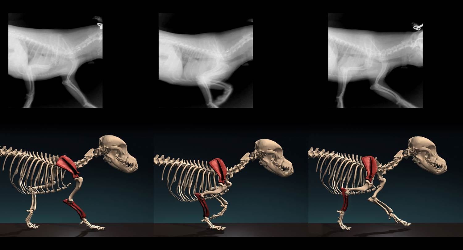
 Einzelbilder aus der Animation der Vorderbeinbewegung eines Chihuahua, erstellt durch den Graphikdesigner Jonas Lauströer
Für die Visualisierung der Struktur-Funktionsbeziehungen im Bewegungssystem werden Knochenmodelle durch hochauflösende Computertomographie generiert und auf der Basis der biplanaren Röntgenaufnahmen animiert. Diese XROMM-Technik („X-ray reconstruction of moving morphology“) wurde von Kollegen der Brown-University entwickelt, die mit uns und anderen Bewegungslaboren einem von der National Science Foundation seit 2009 geförderten Netzwerk zur Koordination röntgenbasierter Bewegungsanalyse angehören. Ziele dieses Netzwerkes sind u.a. die Ausbildung des wissenschaftlichen Nachwuchses, die Entwicklung von spezifischen Methoden und Techniken der Datenerhebung und –auswertung sowie die Etablierung von Datenbanken und Sammlungen für die Röntgenvideographie.
Tierschutz
Unsere Bewegungsstudien an Tieren verschiedener Gruppen der Landwirbeltiere unterliegen geltendem Tierschutzrecht und werden von der zuständigen Ethikkommission des Thüringer Landesamtes für Lebensmittelsicherheit und Verbraucherschutz, Abteilung Veterinärwesen, Pharmazie, gesundheitlicher Verbraucherschutz geprüft. Nur genehmigte Versuchsprojekte werden unter Kontrolle des Tierschutzbeauftragten der Friedrich-Schiller-Universität Jena und des Tierschutzbeauftragten des Institutes durchgeführt. Untersuchungen am Menschen sind grundsätzlich ausgeschlossen.


