Hochgeschwindigkeits-Röntgenvideographieanlage
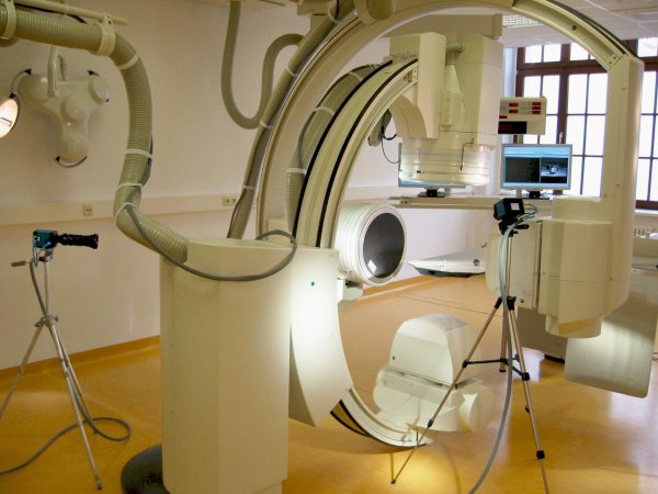
Die biplanare Hochgeschwindigkeits-Röntgenvideographieanlage (Neurostar®, Siemens AG) ist der Prototyp einer umgerüsteten Angiographieanlage. Sie besteht aus zwei Strahlenquellen und zwei zugehörigen Bildverstärkern (Durchmesser: 40 cm), die an zwei C-Bögen befestigt sind. Die beiden Bögen sind getrennt voneinander in nahezu jeder Raumrichtung schwenkbar und können so in verschiedenen Winkeln zueinander eingestellt werden. Der Abstand zwischen Röntgenröhre und Empfänger ist auf minimal 52 cm und maximal 75 cm einstellbar. Die Steuerung erfolgt über eine speziell entwickelte zentrale „intelligente“ Bedieneinheit, die die Röntgenröhren vor Überlastung schützt. Mit ihr können alle für die Röntgenszenen relevanten Parameter in einem großen Variationsbereich vorgewählt werden (Röhrenfokus, Spannung, Stromstärke, Zoom des Bildverstärkers, maximale Szenenzeit, Irisblende). Dadurch lassen sich optimierte Programme für die konkrete Fragestellung und das zu untersuchende Tier zusammenstellen.


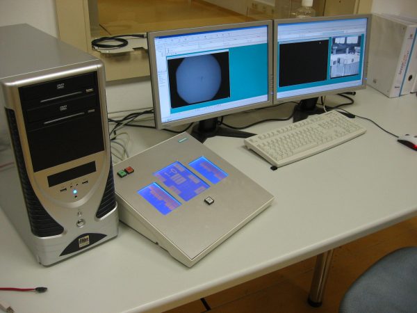
Hochgeschwindigkeits-Röntgenvideographieanlage (biplanarer Aufbau) in Kombination mit zwei zusätzlichen Highspeed-Kameras für simultane Normallichtaufnahmen. Kontrollraum mit zentraler Anlagenbedienung.
Zwei integrierte Highspeed-Kameras (SpeedCam Visario g2, Weinberger GmbH) filmen die Röntgenbilder vom jeweiligen Bildverstärker ab. Die entstehenden digitalen Videosequenzen können in Abhängigkeit von Aufnahmefrequenz und Bildauflösung bis zu 16 Sekunden lang sein. Limitierend dabei ist der Speicher der Kameras. Die Aufnahmefrequenz beträgt 50 - 2000 Hz, die Auflösung ist wählbar von 512 192 über 768 512 und 1024 768 bis 1536 1024 Pixel. Mit Hilfe eines Shutters kann Unschärfen bei sehr schnellen Bewegungen entgegengewirkt werden. Durch die Zoom-Möglichkeit von 40 cm (16 Zoll, Vollbild) über 28 cm (11 Zoll) und 20 cm (8 Zoll) bis zu 14 cm (6 Zoll) können bei genügender Elektronendichte Strukturen bis zu 0,1 mm aufgelöst werden. Vor den Bildverstärkern lässt sich ein Gitter einschieben, das als Referenz zur Korrektur von Verzerrungseffekten dient (Kissenverzerrung, S-Verzeichnung). Die Strahlungsquelle arbeitet mit einer niedrig dosierten kontinuierlichen Strahlung. Besonders bei kleinen Tieren ist vergleichsweise wenig Strahlung nötig, um den Körper zu durchdringen. Zur Einrichtung von Versuchsaufbau und Kameras sowie zur Einstellung der passenden Röntgenparameter gibt es zusätzlich einen Low-Dose-Mode bei einer Aufnahmefrequenz von 100 Hz. Zwei zusätzliche monochrome Normallicht-Highspeed-Kameras (SpeedCamMiniVis, Weinberger GmbH) können frei in der Szene positioniert werden, um einen Überblick zu geben oder ein Detail (z.B. Auffußmoment) zu überwachen. Sie haben eine hohe Lichtempfindlichkeit, eine Auflösung von 512 x 512 Pixeln und eine Aufnahmefrequenz von bis zu 2000 Hz.
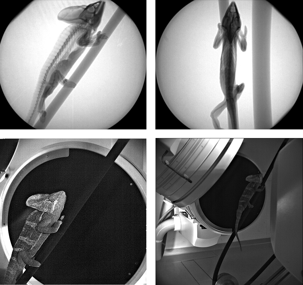
 Synchrone Aufnahme eines Chamäleons mit den vier Kameras. Röntgenaufnahmen von der Seite und von oben, Normallichtaufnahmen als
Detail und Übersicht.
Die Kameras sind synchronisiert und alle vier Videos können gleichzeitig live mit Hilfe der Software Visart (HS Vision) überwacht werden. Durch die Nutzung eines post triggers sind die gespeicherten Aufnahmen synchron. Sie können dann mit Visart weiter bearbeitet werden (Auswahl des relevanten zeitlichen Ausschnittes, Kontrastverstärkung, Schärfefilter etc.). Die Wiedergabefrequenz kann dabei von Einzelbildwiedergabe bis Zeitraffer gewählt werden. Zusätzlich gibt es einen externen Trigger, der die synchrone Aufnahme anderer Daten zu den Bildaufnahmen gestattet, so zum Beispiel Kraftmessdaten, Elektromyogramme, Tonaufnahmen oder Herzdruckmessungen. Der externe Trigger funktioniert über ein 5V Signal. Die Rohdaten werden auf dem Archivserver der Universität Jena gesichert. Je nach weiterer Nutzung können sie in verschiedenen Formaten, beispielsweise als .avi, .tif oder .png gespeichert werden. Die Rohdaten sind nicht entzerrt und nicht kalibriert. Kalibrierkörper und Verzerrungskorrektur-Gitter werden zu jeder Versuchsanordnung gefilmt und liegen ebenfalls als Rohdaten vor.


